To determine the health of the fetus and the degree of its development in gynecology and obstetrics, the study of fetal tissue or amniotic fluid is widely used. To determine the pathology in the baby as early as possible, a study called “chorionic villus biopsy” is carried out for up to 12 weeks. This method is used only after pathology has been identified using ultrasound. At the Other Gynecology clinic, chorionic biopsy is performed in compliance with all rules, so the analysis does not interfere with the development of the baby.
Indications
The chorion represents the future placenta, which consists of the same tissues as the fetus. Cytological analysis of these tissues helps to accurately determine the sex of the child and determine the likelihood of developing intrauterine diseases.
Chorionic biopsy is performed in the following cases:
- the age threshold of the expectant mother is more than 35 years;
- the presence in the family of a child with congenital pathologies;
- consanguineous marriage;
- chromosomal abnormalities in one of the parents;
- the likelihood of developing metabolic disorders or genetic diseases in the baby;
- difficult pregnancy, infections in the first trimester.
The study is also performed when exposed to toxic substances, radiation, viral infections, as well as harmful factors due to the profession of the expectant mother. All this can cause congenital diseases.
Preparation
The likelihood of a baby developing a disease is determined based on predicting the future and analyzing the history of previous pregnancies and pathologies in the parents.
The doctor can make three verdicts:
- high probability (more than 20%) – chorionic villus biopsy must be performed;
- average level of risk (from 5% to 20%) – non-invasive diagnostic methods can be used;
- low risk (less than 5%) – chorionic villus sampling is not necessary.
For the intervention to be successful, it is necessary to prepare. Doctors prescribe the following tests:
- general blood test, urine test;
- gynecological smears and testing them for bacteria;
- blood test for HIV infection, hepatitis, syphilis;
- Ultrasound of the uterus.
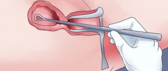
Forceps biopsy technique
The first stage of the intervention is treating the genitals with antiseptic agents to eliminate the risk of infection. Next, the instrument is carefully inserted through the cervical canal into the cavity of the reproductive organ. It consists of thin tweezers that have closed ends. As soon as the instrument approaches the chorion, the ends are opened and a piece of tissue is taken with their help.
The tissue volume must be at least 5 mg. This technique is used for low position of chorionic tissues.
In the article we will consider a research method - chorionic villus biopsy, to determine the health of the baby and identify pathologies in the early stages of pregnancy.